Featured

Welcome to Achieve True Health

This is the post excerpt.


Hello there!

Many of you know I have been struggling with Interstitial Cystitis for almost 10 years. Through my search for an answer, I have learned that all chronic illnesses share 2 things in common: A dysfunctional gut and a dysregulated immune system. To help educate others, I have created a blog. If you know anyone who is struggling with any CHRONIC illness such as IC, Fibromyalgia, Hashimotos, Ulcerative Colitis, Celiac…and the list goes on and on…PLEASE SHARE. The info can save their life!

This is my first post on my new blog.  Honestly, I should have started this blog sooner. If you know what I have been through, you too will wonder why I waited so long to share with you my story.  It has been..well, interesting to say the least.  For those of you who know Carowinds, I equate this with the “Fury”.  Some parts are exciting, but when they are bad, they are downright miserable to the core!

I also have to thank a few people for inspiring me to begin this aggressive journey.  My wonderful partner in crime, Terry, who has been my backbone and support !  He has been a blessing in our lives, and has really pushed me get answers and not just “accept it”, as the doctors would have you.  The other people I have to thank are my team of practitioners: Dr. Bagwell with Maximized Living, Dr. Porter with Novant Health, Bre Walker with Dominion Integrative, and Andrew and Mallorie with On Point Acupuncture!  They have all opened my eyes to a different way of thinking about health and functional medicine.

I don’t want to bore you with too much of my life’s details, but I think it is important that you know how this started and where I am today.

A little about me:  I am 38 years old, divorced, and have 2 beautiful children.  An 8 year old girl and 4 year old boy.  I live in Charlotte, NC.  I am originally from NJ, where I was married and finished college.  We moved here in 2007 to “have a better life”, FWIW.

Fast forward to 2007, and the birth of my first born, via C-section.  About 1.5 to 2 years later, I am afflicted with this condition.  It really came on suddenly. I have always noticed I urinated more frequently, especially during times of high stress.  But the pain came on fast, less than two years after she was born. How can I describe this pain ?  Burning, stabbing, fire, heat, throbbing.  There were times I cried when I had to urinate, and my poor daughter, two years old, would witness my misery.  I felt like I was in an INFERNO.

I immediately went to the gynocologist, figuring this is a UTI. I never had one before, but this is how it feels, right ladies?  Well, to my demise, the lab result was…..NEGATIVE.  What?  How could this be?  Where is this pain coming from.  The doctor proceeds to tell me he is diagnosing me with IC using diagnosis of exclusion, and that I can use a new drug called Elmiron. Ok great doc, gimme the medicine and I see you later.  Well , not so fast.  The catch is, the longer you take this medicine, the better you will feel……………….

What?

Do you mean I have to take this drug forever?

Yes, that is what I am saying.  The drug’s action works better if you take it longer.

Thank you doctor, but no thank you.

I left and did not fill the $500 prescription. Oh and I found out it makes your hair fall out! One thing I know, I have beautiful hair, no way I am going to go bald.

This was the start of my journey.  I would like to call this a journey from hell, but that would be unfair to label that so poorly. I have had some really rough times with this, as you will see in this blog.  But the journey has allowed me to grow in many ways.  It has humbled me at times. It has force me to educate myself, and learn about my body.  Along the process, I continued with my education, and pursued a Masters Degree in Biology. During my studies, I was able to correlate the things i learned in class with my disease.  It has taught me compassion, patience, and prioritization. It has also forced me to appreciate my life. Yes, that means stop and smell the flowers.

I am still on this journey. I am not cured.  But I am getting pretty damn close.

Welcome to my blog!


post

Vitamin D and Your Immune System

vitmainD.immune

The nutrient I wanted to summarize is Vitamin D. I have been fascinated with the role of vitamin D in health topics such as autoimmunity for my own personal reasons. I feel that raising my levels of Vitamin D has been key in my immune system and healing from my autoimmune disease. There is increasing epidemiologic evidence linking vitamin D deficiency and autoimmune diseases including multiple sclerosis (MS), rheumatoid arthritis (RA), diabetes mellitus (DM), inflammatory bowel disease and systemic lupus erythematosus (SLE) (Aranow, 2011).

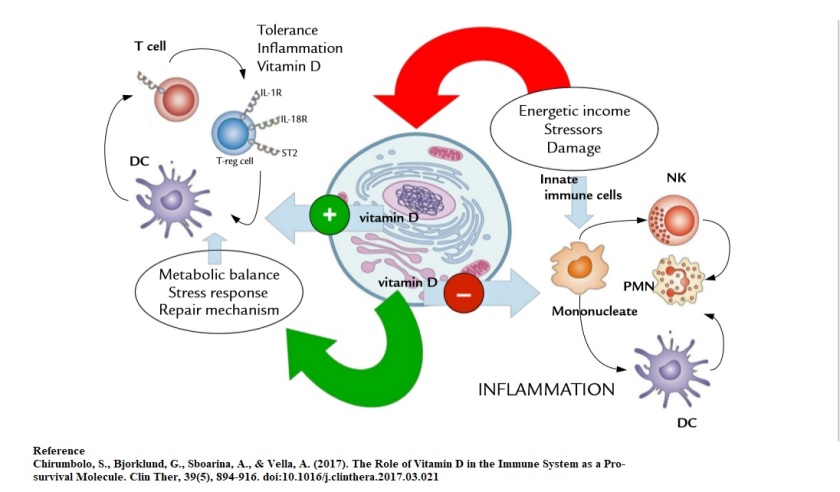
Vitamin D is sometimes called a pro-survival molecule, having anti-inflammatory and immunomodulating action, giving it “stress-quenching activity” (Chirumbolo, Bjorklund, Sboarina, & Vella, 2017).  In fact, research is demonstrating that vitamin D may have the abilities beyond its immune/anti-inflammatory role by inhibiting metabolic stress and energetic expenditure in a cell microenvironment (Chirumbolo et al., 2017). Vitamin D is considered to have a pleiotropic role in the immune system. According to Chirumbolo e al, Vitamin D is involved in the innermost balance of energetics and survival by the cell in its involvement in intracellular organelles of the cell such as the endoplasmic reticulum (ER).  An interesting suggestion comes from evidence of Vitamin D’s involvement in macrophage differentiation and foam cells” (Chirumbolo et al., 2017).

Vitamin D is growing a reputation to enhance human health in at least 4 processes.  Innate immunity, acquired and regulatory Immunity, inflammation, and gut integrity. The area I wanted to dig deeper into was the role of Vitamin D in innate immunity.

Innate Immunity

Researchers are finding that T cells rely on Vitamin D to become activated to detect and kill pathogens.  According to Professor Geisler from the Department of International Health, when a T cell is exposed to a foreign pathogen, it extends the Vitamin D receptor as a signaling device to search for Vitamin D.  “This means that the T cell must have vitamin D or activation of the cell will cease. If the T cells cannot find enough vitamin D in the blood, they won’t even begin to mobilize” (Science Daily, 2010).  T cells that are successfully activated can transform either into killer cells or helper T cells, but they need Vitamin D to perform this successfully.
The activity of vitamin D involves the gut-brain axis, and may play a role in the “hygiene hypothesis” (Chirumbolo et al., 2017).  As a modulatory hormonal molecule, vitamin D can inhibit an inflammatory response and activate regulatory immune mechanisms.  However, it is also “able to induce the production of antibacterial peptides from mononuclear cells and help innate cells to counteract bacterial infection” (Chirumbolo et al., 2017).   In a general way, some scientists consider Vitamin D as an interleukin rather than a “pleiotropic exogenous vitamin”, due to its ability to participate in innate immunity and inflammation, similar to a cytokine.  “Fundamentally, vitamin D dampens the LPS-induced inflammation in mononuclear immune cells (monocytes, macrophages, and DCs) acting on nuclear factor–κB (NF-κB) or p38MAPK via the VDR, which is fundamental to inhibit inflammation” (Chirumbolo et al., 2017).

It was interesting to find that Vitamin D has been previously used to treat infections such as tuberculosis before the era of effective antibiotics, using cod liver oil. In fact, there have been multiple cross sectional studies that associate lower levels of Vitamin D with increased infections.  This is thought to do be due the effects Vitamin D has on the innate immune system.  Vitamin D plays an important role in innate antimicrobial response.  It is thought that when macrophages recognize LPS through TLR, this can increase expression of the vitamin D receptor, and subsequent transcription of antimicrobial peptides (AMP’s) such as cathelicidin and beta defensin (Aranow, 2011).   These peptides show a broad spectrum of antimicrobial activity against bacterial, enveloped viruses and fungi (Kosciuczuk et al., 2012).  AMP’s are super important, since they involve the disintegration of cell membranes of organisms toward which the peptide is active, but do not act on healthy hose cell membranes (Kosciuczuk et al., 2012). Some actions of AMPS’s such as cathelicidins are mediated though their interaction with other cells which are the important part of innate immune system, like monocytes, dendritic cells, T cells and epithelial cells (Kosciuczuk et al., 2012) The susceptibility to bacterial infections of animals and humans with lowered expression of antimicrobial peptides shows their crucial role in the immune response.

Other effects vitamin D has on the immune system is involved in B and T cell mechanisms.  Vitamin D can inhibit B cell proliferation while blocking B cell differentiation and immunoglobulin secretion.  It can also suppress T cell proliferation resulting in subsequent shift from TH1 to TH2 phenotype.  It can also affect T cell maturation with a shifting away from TH17 expression, while facilitating the induction of T regulatory cells.  “These effects result in decreased production of inflammatory cytokines (IL-17, IL-21) with increased production of anti-inflammatory cytokines such as IL-10” (Aranow, 2011).  It can also inhibit monocyte production of inflammatory cytokines such as IL-1, IL-6, IL-8, IL-12 and TNF-a. It additionally inhibits DC differentiation and maturation, as evidenced by a decreased expression of MHC class II molecules, costimulatory molecules and IL-12.  Inhibition of DC differentiation and maturation is particularly important in the context of autoimmunity.

I believe we are just starting to uncover the effects the Vitamin D on the immune system, and it is something I am very interested in learning more deeply.  Some areas to look further include VDR mutations, why some people experience side effects from Vitamin D supplementation, enhancing vitamin D bioavailability from food and gut microbes.  “Given the importance of vitamin D for a functional immune system and the profound deficiency observed in autoimmune disease, as well as the correlation of deficiency with more active disease, an important issue is whether or not the immune components in autoimmune disease are capable of responding appropriately to vitamin D” (Aranow, 2011)

References

Aranow, C. (2011). Vitamin D and the immune system. J Investig Med, 59(6), 881-886. doi:10.2310/JIM.0b013e31821b8755

Chirumbolo, S., Bjorklund, G., Sboarina, A., & Vella, A. (2017). The Role of Vitamin D in the Immune System as a Pro-survival Molecule. Clin Ther, 39(5), 894-916. doi:10.1016/j.clinthera.2017.03.021

Kosciuczuk, E. M., Lisowski, P., Jarczak, J., Strzalkowska, N., Jozwik, A., Horbanczuk, J., . . . Bagnicka, E. (2012). Cathelicidins: family of antimicrobial peptides. A review. Mol Biol Rep, 39(12), 10957-10970. doi:10.1007/s11033-012-1997-x

Science Daily. (2010). Vitamin D crucial to activating immune defenses. Retrieved (2018, June 3) from https://www.sciencedaily.com/releases/2010/03/100307215534.htm

Attachment Parenting and Long Term Stress Response

cryinbaby

I often wondered what impact childhood stress had on a developing child’s brain, particularly during infancy.  When my children were infants, I practiced co-sleeping, baby wearing, and attachment parenting. This created quite a bit of conflict with my ex-husband, who believed in sleep training.  I am glad we are discussing this topic, as it seems that sleep training can be viewed as a type of ACE.

The ‘cry it out’ approach seems to have arisen as a solution to the dissolution of extended family life in the 20th century (Narvaez, 2011).  The idea was, “a baby older than six months “should be taught to sit silently in the crib; otherwise, he might need to be constantly watched and entertained by the mother, a serious waste of time.” According to Narvaez (2011), “we know now that leaving babies to cry is a good way to make a less intelligent, less healthy but more anxious, uncooperative and alienated persons who can pass the same or worse traits on to the next generation”.

I recall during my Masters we studied the experiments on rats, called “Lick your Rats”.  This study was eye opening, as it showed the effects of rats that are nurtured vs. those who were not nurtured.  The researchers indicate that there are hundreds of genes affected by nurturance. In studies of rats with high or low nurturing mothers, there is a critical period for turning on genes and anxiety. “If in the first 10 days of life you have a low nurturing rat mother (the equivalent of the first 6 months of life in a human), the gene never gets turned on and the rat is anxious towards new situations for the rest of its life, unless drugs are administered to alleviate the anxiety (Naravez, 2011)”.  When a baby gets distressed during sleep training, high levels are cortisol are secreted.  It is thought that extreme stress and bouts of high cortisol can negatively influence neural development (Cassels, n.d.).  In cases of severe neglect, effects are seeing the development of white matter in the brain, hippocampus and amygdala. The child can develop a “stress-reactive profile”, similar to the rats in the study, in which they have a heightened response to stress.  Evolutionarily, this makes sense.  An infant raised in an environment in which he or she is not safe has to be acutely aware of the stress around them (Cassels, n.d).

I found it interesting that although babies are typically more hyporesponsive to cortisol and stress, the responsiveness depends on the source of stress.  In fact, separation from the caregiver happens to be one behavior that does elicit a cortisol response, especially if there is no other nurturing caregiver buffering the stress, such as another family member or babysitter.  Essentially, this means that if the baby is left with a non-nurturing caretaker, the negative effects of the separation stress are more impactful.  The behavior or circumstances that elicit cortisol spikes in infancy are moderate-severe pain, abuse, neglect, or abandonment of a caregiver with no responsive substitute.  Sleep training falls well into this category.

Moreover, the infants and their mothers had lost what is called “synchrony”.This is the physiological link between mother and infant, the link that allows mothers to help soothe their babies when upset, the link that is associated with attachment status.

I am happy to say I continued to stand for what I believed in and I coslept with both my children. My daughter was a bit more needy, as I saw immediately in the early days we tried to sleep train.  My motherly instinct took over immediately, and I coslept with her until she was 3 years old.  To this day, she loves when I sleep next to her (at age 10).  My son was a bit more independent, but he coslept for about 2 years.  When I first separated from my ex-husband, we shared a family bed for about a year.  I do not regret a moment of my decision.

“The functional effect of sensitive, responsive, attentive caregiving is that it allows children to express and experience distress, communicate those emotions to caregivers in ways that can elicit help, without stimulating increases in glucocorticoids” (Narvaez, 2011).


References

Cassels, T. (n.d.). “It’s Just a Little Cortisol”: Why Rises in Cortisol Matter to Infant Development. Retrieved (2018, May 29) from http://evolutionaryparenting.com/its-just-a-little-cortisol-why-rises-in-cortisol-matter-to-infant-development/ (Links to an external site.)

Narvaez, D. (2011, December 11).  “Dangers of Crying It Out”.  . Retrieved (2018, May 29) from https://www.psychologytoday.com/us/blog/moral-landscapes/201112/dangers-crying-it-out

Adverse Childhood Events-and YOUR HEALTH

ACE.jpg

This week in Immunology, we are discussing Adverse Childhood Events and the influence it has on the immune system.  This module struck me deeply, as is inspired me to look more deeply at my own health history through the eyes of the functional medicine timeline.  Having an autoimmune disease, I often wondered what the connection with my childhood was with the inflammation that has contributed to my disease.  This information in this module was indeed eye opening.

Numerous studies have demonstrated that childhood adversities have the potential to increase the risk of many diseases that can increase the likelihood of premature mortality, due to increased levels of inflammation (Chen & Lacey, 2018).  As we know, chronic inflammation is associated with increased risk of diseases such as autoimmune disease, cancer, and diabetes (Chen & Lacey, 2018). Having an autoimmune disease myself, I am always looking to dig deeper at my own root causes.  I am not surprised that my childhood experiences could be playing a significant role.  Being born in a first generation Middle Eastern family, there was much pressure put on me as a child in regards to cultural differences that affected me in many ways.  Being repeatedly exposed to adverse childhood experiences (ACE) can affect the human stress regulatory system, which is accompanied by an increase in chronic inflammation.  I recall as early as in my teens having trouble with managing stress.  After I gave birth to my children, I felt as though my adrenals had “crashed” and was diagnosed with adrenal dysfunction at age 31.  Chronic exposures to stress is associated with a dysregulation of the HPA axis and sympathetic adrenocortical axis, which are accompanied by increased inflammation. The chronic inflammation that results is associated with some of the underlying mechanisms of various illnesses, through activation of the immune system. In other worse, dysregulation of the HPA axis, induced by chronic inflammation, put me in a state of immune dysfunction. In FDN, this is called “Metabolic Chaos”.  “The HPA axis is a powerful modulator of inflammatory activity and is in turn modulated by inflammatory processes, as well as being highly responsive to environmental adversities both in childhood and in adulthood” (Baumeister, Akhtar, Ciufolini, Pariante, & Mondelli, 2016).

Several previous studies have reported an association between childhood trauma and increased levels of pro-inflammatory markers, such as the acute phase protein C-reactive protein (CRP), and of the cytokines interleukin-6 (IL-6) and tumor necrosis factor-α (TNF-α). This provides strong evidence that childhood traumatic events significantly impact on the inflammatory immune system, extending their influence into adult health (Baumeister et al., 2016).  It was particularly interesting to note that childhood trauma can even lead to greater methylation of the glucocorticoid receptor (GR), which is correlated with reduced GR function as demonstrated by a negative feedback loop of the HPA axis (Baumeister et al., 2016).  In addition, the increased inflammation can also impair GR function, leading to sustained GR resistance into adulthood.  Moreover, the GR is crucial in regulating TNF-α signaling and TNF-induced cytokine production, as well as conveying protection against TNF-related tissue damage (Baumeister et al., 2016).  It is apparent that dysregulation of the GR has far reaching impacts in regards to modulating inflammation.

Adverse childhood events can also contribute to metabolic syndrome.  Depression is one of the drivers that can contribute to physiological changes associated with metabolic syndrome.  In particular, depression can upregulate inflammatory processes that contribute to atherosclerosis, insulin resistance, and neurodegeneration (Danese et al., 2009).  Depression has been linked to multiple biological abnormalities, including vascular pathologic changes, autonomic function changes, HPA axis dysfunction, and in particular, immune dysfunction. Depression is associated with increased numbers of circulating leukocytes and pro inflammatory cytokines such as interleukin-1 (IL-1), interleukin-2 (IL-2) and interleukin-6 (IL-6). A hypothesis is that there is a positive feedback mechanism between depression and cardiovascular disease, as evidenced by increased levels of CRP in adulthood (which is often used to predict the development of cardiovascular disease).  This is compounded by metabolic abnormalities such as obesity, dyslipidemia, glucose intolerance, and hypertension that exert bi-directional influences on hormone function that lead to hormone imbalances (Danese et al., 2009).  “Many studies reported that the clinical depression can nearly double the risk of mortality and nonfatal cardiac events and that even subclinical elevations in depressive symptoms are related to a poorer prognosis in patients with established CVD” (Liu et al., 2017).

The progression is indeed bi-directional. Increased concentrations of pro inflammatory cytokines influence atherosclerotic plaque progression, poor lifestyle habits and sickness behavior. Poor lifestyle behaviors and sickness behavior can lead to an inactive depressed lifestyle that further heighten the risk of cardiovascular disease (Liu et al., 2017).

In regards to our role as clinical nutritionists, I think this is a great opportunity to education parents on the promotion of healthy psycho-social experiences for children as a potentially cost-effective strategy for the prevention of age-related disease.

References

Baumeister, D., Akhtar, R., Ciufolini, S., Pariante, C. M., & Mondelli, V. (2016). Childhood trauma and adulthood inflammation: a meta-analysis of peripheral C-reactive protein, interleukin-6 and tumour necrosis factor-alpha. Mol Psychiatry, 21(5), 642-649. doi:10.1038/mp.2015.67

Chen, M., & Lacey, R. E. (2018). Adverse childhood experiences and adult inflammation: Findings from the 1958 British birth cohort. Brain Behav Immun, 69, 582-590. doi:10.1016/j.bbi.2018.02.007

Danese, A., Moffitt, T. E., Harrington, H., Milne, B. J., Polanczyk, G., Pariante, C. M., . . . Caspi, A. (2009). Adverse childhood experiences and adult risk factors for age-related disease: depression, inflammation, and clustering of metabolic risk markers. Arch Pediatr Adolesc Med, 163(12), 1135-1143. doi:10.1001/archpediatrics.2009.214

Liu, R. H., Pan, J. Q., Tang, X. E., Li, B., Liu, S. F., & Ma, W. L. (2017). The role of immune abnormality in depression and cardiovascular disease. J Geriatr Cardiol, 14(11), 703-710. doi:10.11909/j.issn.1671-5411.2017.11.006

Allergies and Your Environment

allergies

Allergies are rampant!  I think everyone I know has one type of allergy, whether it skin related, upper respiratory or food!  What is going on?  Why is the prevalence of allergies exploding?  If you want to know more, read below….

Although the term allergy is used loosely to refer to any reaction as an allergy, the medical community defines an allergy as a precise cascade of biochemical reactions. In genetically predisposed (or atopic) individuals, these reactions result in very specific symptoms. Atopy refers to the tendency for asthma, eczema and hay fever, characterized by an overactive immune response to environmental factors (Stanway, 2004).  Symptoms include sneezing, wheezing, bronchoconstriction and even anaphylaxis (Rakel, 2018).

Much research has been focused upon the modification of the allergic immune response early in life, either during the prenatal period or early childhood. Atopy may be genetic, although environmental factors are thought to play a strong role in the development of atopy. Some examples include prenatal and postnatal infection exposure, timing of food introduction, and household pet exposure as predictors of atopy later in life (Rakel, 2018).  The antecedents I would like to focus on are related to environmental factors.

Environmental Factors

Environmental factors interact with genetic factors to maintain allergic (TH2) immune responses, which activate eosinophils, promote IgE production which drive up allergy symptoms (Delves, n.d.).  Interestingly, early childhood exposure to bacterial and viral infections and endotoxins may shift the immune response that can discourage allergic responses (Delves, n.d.).  Trends in developed countries toward smaller families with fewer children and cleaner indoor environments are often thought to contribute to a phenomenon known as the “hygiene hypothesis”. This hypothesis proposes that excess ‘cleanliness’ in an infant’s or child’s environment can lead to a decline in the number of infectious stimuli that are necessary for the proper development of the immune system (Stanway, 2004).  This includes early use of antibiotics that may limit children’s exposure to the infections that drive the opposite immune responses that drive down the allergic reponse, which can explain some of the rise of atopic allergies.  Other factors can include chronic allergen exposure, diet and environmental pollutants.

The hygiene hypothesis also indicates that the early microbial environment is also essential for proper development of protective antimicrobial and regulator immune responses to environmental antigens (Liu, 2015). For example, parents who orally “cleaned” their pacifiers were less likely to have allergic sensitization, eczema and asthma by 18 months (Liu, 2015).  Globally children who have older siblings demonstrate less risks of hay fever and eczema, which were more strongly correlated in more affluent countries (Liu, 2015).  Interestingly, having older siblings may also influence the microbiome.  “The infant gut microbiome associated with having older siblings was recently shown in a birth cohort to have more Bifidobacterium, Lactobacillus, Escherichia, and Bacteroidetes genera and less Clostridia” (Liu, 2015).

In another example, a house dust microbiome that is rich and diverse in the first year of life was negatively associated with allergic sensitization and atopic recurrent wheezing at age 3 years, possibly due to the abundance of Firmicutes and Bacteroidetes phyla.  The same trend has been demonstrated in a household that has dogs.

In laboratory models, oral exposure to extracts of house dust from homes with dogs prevented the development of allergy associated asthma while altering the gut microbiome, including enrichment of Lactobacillus johnsonii (Liu, 2015).

Another area that has been given attention lately is the relationship between allergic diseases and helminth infections. Yes, its a type of worm!  It has been demonstrated that helminths can induce a strong immune response while consequently being inversely associated with allergy and asthma!  These include inducing dentritic cells to induce IL-10 producing Treg lymphocytes while inhibiting allergic airways inflammation in laboratory models of asthma (Liu, 2015).  On the flip side, the innate immune system can also affect immune response against helminth infections which can induce inflammation. “This reaction is the body’s response to destroy helminths, especially tissue helminths that stimulate an excessive immune response that can promote allergies. Though inconclusive, it does demonstrate the role the microbial environment has on atopic allergic responses.


References

Delves, P. (n.d.) Overview of Allergic and Atopic Disorders. Retrieved (2018, May 22) from https://www.merckmanuals.com/professional/immunology-allergic-disorders/allergic,-autoimmune,-and-other-hypersensitivity-disorders/overview-of-allergic-and-atopic-disorders

Liu, A. (2015). Revisting the hygiene hypothesis for allergy and asthma. J Allergy Clin Immunol 2015;136:860-5.

Rakel, D. (2018). Integrative Mecidine (Vol. 4): Elsevier Inc.


Sitcharungsi, R., & Sirivichayakul, C. (2013). Allergic diseases and helminth infections. Pathog Glob Health, 107(3), 110-115. doi:10.1179/2047773213y.0000000080

Stanway, A.  (2004). Causes of atopic dermatitis. Retrieved (2018, May 22) from https://www.dermnetnz.org/topics/causes-of-atopic-dermatitis/


Mast Cell Activation and Inflammation

strawberries

I love strawberries!  They are high in Vitamin C and just delicious on salads.  Sadly, they also can cause pain in many people who have mast cell activation disorder. Read below….

I have recently become very interested in histamine intolerance and mast cells, and strawberries is on the list of foods high in histamine. There is some significant research being done on mast cell activation, autoimmune disease and the immune system.  Histamine intolerance is the disequilibrium between accumulated histamine and histamine degradation (Manzotti, Breda, Di Gioacchino, & Burastero, 2016).  Histamine degradation is dependent on the primary enzyme, diamine oxidase (DAO). Histamine is a biogenic amine found in foods such as pickles, matured cheese, fermented foods and leftovers.  Some foods are histamine liberators and that includes fruits such as pineapples, bananas, citrus fruits, papayas and strawberries.  The ingestion of histamine rich foods can provoke a variety of symptoms such as digestive, arrhythmia, flushing, asthma, hypotension, rhinoconjunctivitis, and headaches.  Impaired DAO production is often the culprit, which can result in increased enteral histamine uptake and increased plasma histamine concentrations (Manzotti et al., 2016).  Interesting the study by Manzotti demonstrated that 71% of patients reported functional bloating after consuming high histamine foods.

The latest research indicates that mast cells that release histamine and other inflammation in the bloodstream are active participants in autoimmune disease related tissue damage.  Increased mast cell activity, release of histamine and other inflammatory agents are frequently seen in autoimmune conditions such as MS, RA and many others (Healing Histamine, n.d.).   A variety of receptors including those engaged by antibody, complement, pathogens, and intrinsic danger signals are implicated in mast cell activation in disease.  A variety of receptors including those engaged by antibody, complement, pathogens, and intrinsic danger signals are implicated in mast cell activation in disease (Brown & Hatfield, 2012).  Mast cells can also recruit other immune cells such as neutrophils, to the sites of autoimmune destruction.  “Mast cells can only act as accessory cells to the self-reactive T and/or antibody driven autoimmune responses” (Brown & Hatfield, 2012).  This includes mast cells among one of the major contributors to autoimmunity and should definitely be considered.

Autoimmune and allergic diseases share fundamentally important features in that both are the result of “hypersensitive” immune responses directed toward inherently harmless antigens (Brown & Hatfield, 2012).  An early progress of autoimmune disease involves the activation and expansion of T and/or antibody producing B cells that wear autoreactive receptors.  These autoreactive T or B cells can then enter the bloodstream and migrate to sites of inflamed tissues expressing relevant autoantigens (Brown & Hatfield, 2012). “T cells, through the elaboration of cytotoxic mediators, and antibodies, through complement fixation or their ability to activate resident accessory cells such as macrophages and mast cells via Fc receptor engagement, can play direct roles in tissue destruction at these sites” (Brown & Hatfield, 2012)

In addition to eliciting the above described adaptive immune response, antigens can engage a class of danger- associated receptors on both adaptive and innate immune cells, including mast cells.  These receptors include toll-like receptors (TLRs) and Nacht-LRRs (NLRs) (Brown & Hatfield, 2012).  Activation of immune cells through TLRs and NLRs induces the expression of multiple inflammatory mediators that can ultimately result in local tissue damage causing the release of normally sequestered tissue antigen (Brown & Hatfield, 2012). Subsequent recognition of these antigens by T or B cells will induce activation and initiate autoimmunity. Alternatively, infection-induced activation of accessory immune cells, including mast cells, macrophages, and neutrophils, can boost inflammation and transform a relatively modest autoreactive response.

There is evidence that other TLRs are expressed on mast cells.   For example, activation of mature mast cells through TLR2 results in their production of several pro-inflammatory cytokines critical in autoimmunity including IL-17, IFNγ, TNF, and IL-1β.  Mast cells also express multiple IgG Fc receptors. This is significant because IgG autoantibodies are hallmarks of many autoimmune diseases and have been detected in multiple autoimmune diseases.


References

Brown, M. A., & Hatfield, J. K. (2012). Mast Cells are Important Modifiers of Autoimmune Disease: With so Much Evidence, Why is There Still Controversy? Front Immunol, 3, 147. doi:10.3389/fimmu.2012.00147


Healing Histamine (n.d.).  Histamine Intolerance, Mast Cells & Autoimmunity.  Retrieved (2018, May 15) from https://healinghistamine.com/histamine-mast-cells-autoimmune-disorders/


Manzotti, G., Breda, D., Di Gioacchino, M., & Burastero, S. E. (2016). Serum diamine oxidase activity in patients with histamine intolerance. Int J Immunopathol Pharmacol, 29(1), 105-111. doi:10.1177/0394632015617170

Erectile Dysfunction and Inflammation

viagra-pills

Cytokines, are small secreted proteins released by cells have a specific effect on the interactions and communications between cells. Different cytokines are associated with different diseases or conditions(Zhang & An, 2007). One particular cytokine, TNF-α, has been shown to play an important role in ED as well as cardiovascular disease (CVD), due to the effect it has on the vascular system.  Patients with ED often show high levels of TNF-a, which is a cytokine that is often secreted by white blood cells in response to inflammatory stimuli.  In addition, the presence of a low-grade inflammatory process is associated with many cardiovascular diseases (CVD). Cytokines levels, such as TNF-α, are increased in response to inflammation and contribute to the changes in vascular reactivity observed in CVD (Carneiro et al., 2010). Two distinct surface receptors mediate the effects of TNF-α. These include TNFR-1, and TNFR-2.  Gene expression of TNFR-1 has been demonstrated in cavernosal tissue of the penis (Carneiro et al., 2010).  It has been pretty well established that this cytokine is an important contributor of many cardiovascular diseases related to endothelial dysfunction. Additionally, endothelial function is impaired in inflammatory conditions and conditions with increased oxidative stress.  In vivo laboratory administration of TNF-α demonstrates impairment of endothelium-dependent vasorelaxation in a variety of vascular beds and decreases the release of nitric oxide (NO), which is required for erectile function. TNF-α has the ability to increase arterial reactive oxygen species generation, which likely accounts for some of the reduction in NO levels.

So the question is, what is causing the TNF-α to be elevated?  I found the link to environmental toxins to be quite intriguing.  Could exposure to these toxins induce elevations in inflammatory cytokines, like TNF-α, that leads to downstream effects such as vascular dysfunction?  Some environmental toxins, such as endocrine disrupting chemicals (EDCs) interfere with the synthesis of cytokines, immunoglobulins, and inflammatory mediators, and they also affect the activation and survival of immune cells (Yang et al., 2014).  The dysfunction of the immune system caused by some EDCs may lead to immunity immunodeficiency against infection, or to hyperreactivity of immune responses, as seen in allergy and autoimmune disease (Yang et al., 2014).  Nonylphenol (NP), one of the alkylphenols, is the most important metabolite of a group of nonionic surfactants, is a common EDC that can bioaccumulate through the diet.  It is structurally similar to estrogen, which can feminize male animals and may be linked to infertility.  Some in vitro or ex vivo studies suggest that NP skews T cells towards Th2 responses through its influence on dendritic cells (DCs), resulting in increased expression of IL-6 and TNF-α, but not IL-10 and IL-12, in response to LPS stimulationNP increases the expression of TNF-α, but suppressed anti-inflammatory cytokine IL-10 production in a range of physiological doses, burning both ends of the candle.

Another interesting association with increased levels of TNF-α is associated with intestinal parasites such as Entamoeba histolytica.  Although TNF can be synthesized and secreted by many other cell types, such as neutrophils, eosinophils, and mast cells, the main source of TNF is activated macrophages.  Usually, TNF contributes to the control of parasitic and bacterial infection like in the case of Trypanosoma infection or by triggering an immune response. E. histolytica activates the inflammatory process by promoting TNF production, which is thought to be crucial for guiding E. histolytica towards TNF-producing cells of the host. One may speculate that this behavior is triggered by its need of nutrients that cannot be satisfied in the inflamed tissues. Inflammation causes change in the gastrointestinal microbiota, and these changes may provoke E. histolytica to search for alternative sources of nutrients.

Other intestinal parasites have been associated with HPA axis dysfunction, since the immune and neuroendocrine systems are interconnected by a network in which hormones, antigens, receptors, cytokines, antibodies and neuropeptides modulate immune response. This indicates that intestinal parasites can influence sex hormone production, such as testosterone.  Low testosterone levels often go hand in hand with erectile dysfunction, but that will be discussed in a future blog.

The bottom line; get to the bottom of the problem. Find the source of the inflammation that is ruining your sex life. Run functional labs such as a stool test to rule out intestinal parasites.  An HPA axis test, such as the Dutch Complete, can determine the hormone imbalance and potentially identify the root cause. An environmental toxin test can reveal if toxins are ruining contributing to your immune dysfunction.  Running these labs will allow you to get on a healing path so you can get off the purple pill and regain your life back. You should work with a practitioner who is knowledgeable about the tests and can work with you to find your path to wellness.


References

Ankri, S. (2015). Entamoeba histolytica – tumor necrosis factor: a fatal attraction. Microb Cell, 2(7), 216-218. doi:10.15698/mic2015.07.216

Carneiro, F. S., Webb, R. C., & Tostes, R. C. (2010). Emerging role for TNF-alpha in erectile dysfunction. J Sex Med, 7(12), 3823-3834. doi:10.1111/j.1743-6109.2010.01762.x

Yang, S. N., Hsieh, C. C., Kuo, H. F., Lee, M. S., Huang, M. Y., Kuo, C. H., & Hung, C. H. (2014). The effects of environmental toxins on allergic inflammation. Allergy Asthma Immunol Res, 6(6), 478-484. doi:10.4168/aair.2014.6.6.478

Zhang, J. M., & An, J. (2007). Cytokines, inflammation, and pain. Int Anesthesiol Clin, 45(2), 27-37. doi:10.1097/AIA.0b013e318034194e

 Reply

Microbiome & Immune Connection

The-human-microbiome-and-human-health.jpg

Microbiome and the Influence on the Immune System-it’s more important than you think!

The composition of the microbiome can influence the function of your IMMUNE SYSTEM.  For example, numerous studies indicate that your microbiome can influence  your susceptibility to certain bacteria such as Campylobacter, which is often seen in anxiety disorders.  In fact, a book by Martin Blaser called “Missing Microbes” discusses that the alteration of the human microbiome could be one of the factors attributed to the rise of conditions such as food allergies, asthma, celiac disease and intestinal disorders such as IBD.  His theory is that the modern medical practices, that includes overuse of antibiotics, may have negatively shifted the microbiome in a way that is affecting human health detrimentally.  The use of antimicrobial drugs is known to be associated with the development of certain infectious diseases, such as colitis and candida overgrowth, through disruption of this delicate microbiome balance (Casadevall and Pirofski, 2018).

It is hypothesized that the establishment of your microbiome occurs during the first 3 years of your life, and disruptions in this establishment during this time can have long term implications (Blaser, 2016).  According to Blaser, 50% of babies in the US are born by C-section, and this can be the beginning of some of the various disruptions in the delicate microbiome balance. This is not limited to bacteria, but also fungal and viral elements of the microbiome, that are also demonstrating a role in human health as well.

Nutritional intervention: Probiotics
A nutritional intervention to address the microbiome is the use of probiotics.  Probiotics are live organisms that, when ingested in adequate quantities, exert a health benefit on the host.  Probiotics can directly stimulate the growth of beneficial bacteria and competitively exclude the number of more harmful, toxin producing microbiota (Zhou & Foster, 2015).  Lactobacillus, Bifidobacterium, and Saccharomyces are three extensively studied and commonly used probiotics in humans and animals.

Probiotics are demonstrating to directly affect your gut immune system, which is responsible for

Three main beneficial effects of probiotics on your gut defense system include the following:

  1. Probiotics can block pathogenic bacteria by producing substances that directly compete with pathogens for adherence to the intestinal epithelium.
  2. Probiotics can enhance intestinal barrier function and stimulate protective responses from epithelial cells. This can lower the chance of bacteria translocating into the blood stream and entering systemically. This function may decrease infections and immune related reactions, thus supporting the health of the immune system (Enviormedica, n.d).
  3. Probiotics are able to modulate the immune system and pathogen induced inflammation through various immune regulated signaling pathways.

Immune Cells

Probiotics can regulate host innate and adaptive immune responses by modulating the functions of your very immune cells.    For example, a probiotic mixture consisting of L. acidophilus, L. casei, L. reuteri, B. bifidium, and Streptococcus thermophilus stimulated regulatory proteins involved in your immune system that are responsible for both inflammatory and anti-inflammatory mechanisms.

Intestinal Epithelial Cells

Probiotics can repair damaged epithelial barrier while producing antibacterial substances and protective proteins, as well as regulate intestinal epithelial immune function (such as cytokine production) (Yan & Polk, 2011).  For example, l. johnonsii has the ability to upregulate various receptors on your immune cells,  which may indicate that the probiotic is able to stimulate the gut immunity. “These findings suggest that probiotics regulation of innate immunity in intestinal epithelial cells may serve as a mechanism for disease prevention and treatment” (Yan & Polk, 2011).

References:

American Society for Microbiology. (2016, September 6).  Missing Microbes with Dr. Martin Blaser.  Retrieved 2018, May 4 from https://www.youtube.com/watch?v=KwK_O0ahDKo

Environmedica. (n.d.). Probiotics for Immune System Support. Retrieved 2018, May 3 from https://www.enviromedica.com/probiotics-immune-system (Links to an external site.)Links to an external site.

Yan, F., & Polk, D. B. (2011). Probiotics and immune health. Curr Opin Gastroenterol, 27(6), 496-501. doi:10.1097/MOG.0b013e32834baa4d

Zhou, L., & Foster, J. A. (2015). Psychobiotics and the gut-brain axis: in the pursuit of happiness. Neuropsychiatr Dis Treat, 11, 715-723. doi:10.2147/ndt.S61997

Endocrine Disruptors and your Health

endocrine.disrupturs.png

Our world is toxic, and lately endocrine disruptors have been the focus of the health industry.  New products are popping all over the market, and may new companies are launching in the market, claiming to be free of endocrine disrupting chemicals.

I just encountered a new product line called Poofy Organics that looks promising with products that are clean and free of these dangerous and pesky chemicals.  They have a diverse product line and best of all, they are affordable! Read on about what these chemicals are, what they do to your body and I will share the link to where you can order if you wanted to try for yourself.

I first encountered the topic of endocrine disruptors from one of my friends a few years ago who started making her own hair and skin care products using cleaner, phthalate free ingredients.  At the time, I did not know much about endocrine disruptors, but it was also around the time that BPA was being removed from baby bottles.

What are endocrine disruptors?

They are chemicals that may interfere with the body’s endocrine system and are showing to have adverse developmental, reproductive, neurological, and immune effects in both humans and wildlife (Endocrine Disruptors, n.d.).  They can be found in a wide range of every day products such as pharmaceuticals, pesticides, plastics, metal food cans, detergents, toys, and cosmetics.  Currently there are numerous studies, supported by the NIEHS, to determine if exposure to endocrine disruptors may result in human health effects, particularly infertility and cancer (Costa, Spritzer, Hohl, & Bachega, 2014).  Studies indicate that endocrine disruptors can affect multiple areas of reproduction, development and metabolism.

Endocrine disruptors primarily exert their effects by binding to hormone receptors and transcriptional factors. (Costa et al., 2014)  There are 3 main methods that affect the hormone levels in the body (Endocrine Disruptors, n.d.):

  1. They can mimic naturally occurring hormones such as estrogens, androgens, and thyroid hormones, potentially over-stimulating (called the agonistic effect)
  2. They can bind to a receptor within a cell and block the endogenous hormone from binding, such as anti-estrogens and anti-androgens (called the antagonistic effect)
  3. They can interfere with the way natural hormones or their receptors are made or controlled, by altering metabolism in the liver.

However, recent studies are showing that they can also alter the enzymes involved in the synthesis or catabolism of steroids (Costa et al., 2014).  I find most alarming is that the highest incidence of reproductive disorders that were observed in the second generation of the women exposed to endocrine disruptors, such as diethylstilbestrol (DES).  This suggests that the epigenetic changes can affect subsequent generations.

Where are endocrine disruptors found?  Everywhere!  They are ubiquitous in our modern society, unfortunately.  Research indicates many places where they can be found to accumulate. There are 3 main categories that they are listed in, which are BPA, DEHP, and phytoestrogens.  However, I felt the EWG did a fairly good job summarizing them down to the Deadly Dozen.   I will summarize 5 of them below:

  1. BPA-biphenol A-It is one of the chemical compounds of highest production worldwide; more than 6 billion pounds are produced annually. (Costa et al., 2014) . It has been identified in 95% of urine samples of the American population and also found in serum of pregnant women. BPA is most recognized on its effects on thyroid hormones, particularly its estrogenic and adipogenic activities, which could definitely linked to metabolic diseases such as hypothyroidism, diabetes, obesity, and cardiovascular disease.  One way to minimize exposure is to eat fresh foods and avoid canned foods.  Also I found it interesting that BPA is found in cashier receipts.  In fact, levels of BPA in cashiers who handle receipts were higher after their shifts than in between shifts. (Our Chemical Lives, 2015)
  2. Phthalates-These are plasticizers used as softeners, but are also found in paints, solvents, toys, personal and medical care products, and cosmetics and blood transfusion bags (Costa et al., 2014) The main routes of human exposure are food and skin absorption, and it can occur during trans placental route and during breastfeeding.  DEHP, Di (2-ethylhexylphthalate), are implicated in the toxicity of the reproductive system and also increased proliferation of fat cells, which predisposes you to visceral obesity (Costa et al., 2014).  Also epidemiological studies have correlated phthalates in cord blood and lower gestational age at delivery. Other areas where studies link their effects in with male fertility, lower sperm counts and defects in male reproductive system  (Deadly Dozen Endocrine Disruptors, 2013). Exposure to DEHP can alter DNA methylation patterns in both the prostate and testicular cells (Sweeney, Hasan, Soto, & Sonnenschein, 2015).  These epigenetic patterns seen are also heritable, meaning DNA methylation patterns and histone modifications have been observed in successive generations (Sweeney et al., 2015).  “Interruption of hormonal function within the developing tissues have been shown to lead anatomical changes in new born male babies as well (Sweeney et al., 2015).
  3. Dioxins-These compromise a group of organochlorine compounds that include PCBs (polychlorinated biphenyls). They are found in insulators, flame-retardants, lubricants, and machine and transformer fluids.  They can disrupt both male and female sex hormone signaling, and if exposed in the womb, they can permanently affect sperm quality and lower the sperm count in men  (Deadly Dozen, 2013).  They are fat soluble and can easily contaminate the food chain because they accumulate in fat cells. They are also not readily metabolized and excreted and half a half-life of 8 years (Costa et al., 2014).  They are found in products like meat, fish, milk, eggs and butter, so the best wait to limit exposure is to cut back on eating animal products.
  4. Pesticides- A recent study in UK reported there are approximately 127 pesticides identified with endocrine disrupting activity. Among the classes of pesticides that stand out are organophosphates, carbamates, and organochlorines (Costa et al., 2014).  Also, there is still some contamination of DDT found in some developed countries despite being banned.  Atrazine is the most commonly detected pesticide found in ground and surface water (Costa et al., 2014).  It is linked to breast tumors, delayed puberty and prostate inflammation in animals, and even prostate cancer in humans (Deadly Dozen, 2013).  Organophosphates can effect brain development, behavior and fertility. They can also interfere with testosterone communication, lowering both testosterone and thyroid hormone levels.  Ways to limit exposure is to buy organic, use EWG’s Shoppers guide to pesticides in produce to assist in finding fruits and vegetables with fewest pesticide residues.
  5. PFC-perflourinated chemicals used in non-stick cookware. I chose this one because I love to cook, but accumulation of this chemical makes home cooked meals not as healthy as they should be! Perfluorochemicals are so widespread and extraordinarily persistent that 99 percent of Americans have these chemicals in their bodies (Deadly Dozen, 2013).  One particular compound, PFOA, has been shown to be resistant to biodegradation, which mean it will never break down in the environment. PFOA is linked to decreased sperm quality, low birth weight, kidney disease, thyroid disease, cholesterol, etc.  It seems to affect the thyroid and sex hormones the most.  One way to minimize exposure is to eliminate the usage of non-stick pans.  There is a pan called the “Green Pan” that is non-stick, ceramic based pan that is made from a sand derivative that does not use PFOA or PFAS during the production process.  It also is heat resistant to temperatures up to 450C and it will not release any toxic fumes if you accidentally overheat it.

Here is some information about the Green Pan:

https://www.greenpan.us/why-ceramic (Links to an external site.)

Here is the link to the EWG 2017Shopper’s Guide to Pesticides in Produce

https://www.ewg.org/foodnews/#.Wfy0prpFxfw (Links to an external site.)

One thing I found was interesting was that licorice (Glycyrrhiza glabra) root extract (LRE) may help alleviate the toxicity of endocrine disruptors (Chu, de la Cruz, Hwang, & Hong, 2014) IT is commonly called “gamcho” in Korea exhibits anti-oxidative, chemo-protective, and detoxifying properties.  In recent studies, special attention has been given to the study of plant and their isolates for the prevention of diseases.  Licorice root has been used in over 70% of Chinese medicines and has been used by human beings for over 4000 years.   According to a study in 2014, there is some promising evidence that licorice root extract can be used as a potential toxicity-alleviating agent to prevent the occurrence of endocrine disrupting-induced tumors and can help modulate the risk of cancer to excessive exposure to endocrine disruptors. Furthermore, licorice root has other healing properties and can act as an adaptogenic agent for modulating adrenal insufficiency, and is definitely worth exploring further.

So for the best part, here is the link to order Poofy.  POOFY ORGANICS was founded in 2006 after a family member was diagnosed with breast cancer.  The family was determined to stop using products laden with toxic chemicals. With no suitable alternatives to turn to, they made it their mission to find the safest and most effective ingredients for our products.  I am waiting for a box of samples to arrive, I can’t wait to try these products out!!!

Poofy logo

https://dena.poofyorganics.com/

References

Chu, X. T., de la Cruz, J., Hwang, S. G., & Hong, H. (2014). Tumorigenic effects of endocrine-disrupting chemicals are alleviated by licorice (Glycyrrhiza glabra) root extract through suppression of AhR expression in mammalian cells. Asian Pac J Cancer Prev, 15(12), 4809-4813.


Costa, E. M., Spritzer, P. M., Hohl, A., & Bachega, T. A. (2014). Effects of endocrine disruptors in the development of the female reproductive tract. Arq Bras Endocrinol Metabol, 58(2), 153-161.

Dirty Dozen Endocrine Disruptors. (2013, October 28). Retrieved from https://www.ewg.org/research/dirty-dozen-list-endocrine-disruptors#.WfykCrpFxfw

Endocrine Disruptors.(n.d.)  Retrieved from http://www.ewg.org/research/dirty-dozen-list-endocrine-disruptors (Links to an external site.)Links to an external site. (Links to an external site.)

Our Chemical Lives. (2015, March 31).  Retrieved from https://www.youtube.com/watch?time_continue=1553&v=J9SWBAUlAvw

Sweeney, M. F., Hasan, N., Soto, A. M., & Sonnenschein, C. (2015). Environmental endocrine disruptors: Effects on the human male reproductive system. Rev Endocr Metab Disord, 16(4), 341-357. doi:10.1007/s11154-016-9337-4


Exercise and Inflammation

fitness.image

Exercise and Inflammation-it’s more than just vanity!

Having worked in the fitness industry for over 15 years and helping people with their weight loss goals, I always wondered what role exercise had on inflammation.  In obesity, various mechanisms are thought to contribute to a low grade inflammation within the fat tissue affecting the development of several secondary diseases such as metabolic syndrome, insulin resistance (IR), diabetes, arterial hypertension and asthma (Schmidt et al., 2015).

The study I examined look at levels role of exercise and how it influences the levels of pro-inflammatory cytokines in participants with high vs. low physical activity (Gleeson et al., 2011).  The study demonstrated a strong relationship to BMI that may indicate that the decrease in inflammatory molecules may be related to decrease in visceral fat.  Interestingly, calorie restriction and fasting has been shown to reduce visceral fat in numerous other studies, as well as subsequent reduction in inflammation (which is an area I am very interested in digging into more).  Additionally, physical activity may further mitigate inflammation by improving endothelial function, increasing insulin sensitivity, enhancing liver health and increasing blood vessel growth and blood flow.

An article by Gleeson et. al discusses the anti-inflammatory effects of exercise and explains 3 main mechanisms of action:

  1. Reduction in visceral mass-as mentioned early, a reduction in visceral mass has an indirect effect of being able to decrease inflammation, since accumulation of fat in the omentum, liver and muscles, as well as the expansion of adipose tissue, results in enhanced production of certain inflammatory mediators. Therefore loss of visceral fat can result in reduction in inflammation.
  2. Release of IL-6 from working muscles-A fall in muscle glycogen content with exercise signals the muscles to secrete IL-6 (a pro-inflammatory cytokine), which stays high during the duration of exercise. However, this also initiates a rise in anti-inflammatory cytokines IL-10 and IL-1RA to minimize the effects on the tissue.  Also, it was interesting that you really need 2.5 hours or more of strenuous exercise to get a significant elevation of IL-6, which may partially explain why marathon runners may have suppressed immune systems.
  3. Increased levels of cortisol and adrenaline-IL-6 stimulates the release of cortisol, which is smaller doses, can have anti-inflammatory effects. It should be pointed out that too much cortisol secretion from the adrenal glands can create a chronic state of inflammation as well, so this could also be a dose dependent phenomenon.

There also appears to be a strong relationship between exhaustive exercise, such as marathon running, and chronic low-grade inflammation induced by the massive systemic release of several pro-inflammatory cytokines and chemokines, such as IL6 and IL8, G-CSF, although host tissue damage may be restricted by compensatory mechanisms.  In conclusion, benefits of regular exercise and physical activity are well observed, but I think more clear direction is needed on the types and intensities as well as the health of the person doing the exercise to understand the impact it may have on inflammation.

References

Gleeson, M., Bishop, N. C., Stensel, D. J., Lindley, M. R., Mastana, S. S., & Nimmo, M. A. (2011). The anti-inflammatory effects of exercise: mechanisms and implications for the prevention and treatment of disease. Nat Rev Immunol, 11(9), 607-615. doi:10.1038/nri3041

Part 2 of 2: Inflammation and Exercise: friend or foe?  (2011, August 25).  Retrieved 2018, May 2 from https://inscientioveritas.org/inflammation-and-exercise/ (Links to an external site.)

Schmidt, F. M., Weschenfelder, J., Sander, C., Minkwitz, J., Thormann, J., Chittka, T., . . . Himmerich, H. (2015). Inflammatory cytokines in general and central obesity and modulating effects of physical activity. PLoS ONE, 10(3), e0121971. doi:10.1371/journal.pone.0121971

IC and Histamine

allergy

I have been very interested in histamine and histamine intolerance from the time I was first diagnosed with IC and noticed that allergy medications helped me during flare ups.  It is well known that there is an involvement of mast cells and cystitis related pain. “Interstitial cystitis has been found in combination with some allergic or autoimmune disorders and histopathological abnormalities resembling allergic disorders, including mast cell activation, histamine release and eosinophil infiltration” (Lee, Doggweiler-Wiygul, Kim, Hill, & Yoo, 2006).  Clinical studies demonstrate elevated mast cell numbers in the lamina propria of IC bladder biopsies.  This may explain why neuromodulatory therapies suggests are partially effective in IC therapies, as “neural-immune interactions play a role in IC pathogenesis” (Rudick, Bryce, Guichelaar, Berry, & Klumpp, 2008).  Mast cells contain preformed stores of immune mediators, such as histamine and TNF, and can be activated to release these stores by neurotransmitters such as substance P, a polypeptide thought to be involved in the synaptic transmission of pain and other nerve impulses. It is hypothesized that that activation of bladder related circuits in the CNS initiate substance P by peripheral nerves in the bladder can promote mast cell activation, mediated by substance P (Rudick et al., 2008).  The mast cells are thought to induce bladder inflammation by acting on the bladder epithelium (lining). Also, increased levels of urinary histamine metabolites have been detected in IC patients.  This is thought to be due to accumulation of mast cells in the lamina propria.

A role for histamine and histamine receptors in pain responses has been documented in both humans and animal models.  “However, histamine can be derived from various cell types, including mast cells, basophils, and histaminergic neurons” (Rudick et al., 2008). More recently, inflammatory cells such as neutrophils and dendritic cells have been shown to produce histamine.  In fact, it has been demonstrated in various clinical settings that antihistamine therapy improves IC-related pelvic pain, and the mechanism for this effect is unknown. However, it can be inferred that this is due to histamine activation.  Irritable bowel syndrome (IBS) is another chronic condition that resembles IC in the characteristics of pathology. “In IBS, pain is correlated with activated colonic mucosal mast cells in proximity to nerves” (Rudick et al., 2008). In fact in IBS, colonic tissue extracts often reveal elevated histamine.  These findings support the idea that mast cell histamine may mediate pain in multiple pelvic pain syndromes (Rudick et al., 2008).

With my clients, we have a test I run by Dunwoody that can test for histamine intolerance. This test will measure hitamine levels, DAO (an enzyme that breaks down histamine), and DAO: histamine ratio. Classic symptoms of histamine intolerance include gas, high blood pressure, arrhythmia, nausea, hives, stomach ache, itching, cramps, headache, sneezing, dizziness, congestion, runny nose, shortness of breath, especially after meals containing histamine.  Low DAO is also associated with the same symptoms along with inflammation, arthritis and certain neurological conditions such as MS.  Low DAO is also associated with leaky gut and poor gut function and immunity.  It can also be a trigger for depression and anxiety.  Individuals with inability to break down histamine seem to “react to everyting” or seem to improve on anti-histamines.   I have to mention that the low histamine diet, combined with DAO enzyme before meals, has been shown to be really helpful in managing IC pain related to mast cells.  To really get on the road to healing and run some of the key functional tests, contact me at mandy@naturalhealthachiever.com so I can better guide you!

References:

Lee, J., Doggweiler-Wiygul, R., Kim, S., Hill, B. D., & Yoo, T. J. (2006). Is interstitial cystitis an allergic disorder?: A case of interstitial cystitis treated successfully with anti-IgE. Int J Urol, 13(5), 631-634. doi:10.1111/j.1442-2042.2006.01373.x

Rudick, C. N., Bryce, P. J., Guichelaar, L. A., Berry, R. E., & Klumpp, D. J. (2008). Mast cell-derived histamine mediates cystitis pain. PLoS ONE, 3(5), e2096. doi:10.1371/journal.pone.0002096